In the past years, different research projects have been conducted in an effort to find a more efficient and accurate way of carrying out the clinical pathway for prostate cancer (PCa) diagnosis.
Thanks to the results of the PROMIS study, reported by The Lancet in 20171, which demonstrated how beneficial including a prostate MRI can be in the diagnostic process of PCa, organizations such as the EAU (European Association of Urology)2 and the ACR (American College of Radiology)3 changed their guidelines for PCa diagnosis to include the acquisition of an MRI pre-biopsy as a standard procedure.
Naturally, the number of prostate MRIs expected to be performed every year will exponentially increase. Which brings the radiology community to a crossroads: is it possible to maintain the quality of patient care taking into account this increase of workload?
The answer is yes, with the right strategy. The following blog will detail what challenges occur together with the increase in workload and how an Artificial Intelligence (AI) software solution for reading prostate MRI can help radiologists overcome the most frequent pain points in the field.
Prostate MRI and the radiologist’s challenges
One of the biggest struggles for professionals in the prostate field is how time-consuming the whole process is, from logging in to all required software for the reading process, to the assessment itself, dictating and report creation. Guidelines are available, however, the standardized process includes a long list of steps, which can be tedious and demanding.
Additionally, many software programs that are available for MRI scan assessment do not offer all functionalities needed for precise evaluation of prostate MRIs. For example, the user would have to use a PACS viewer to look at the images, use an online calculation tool to determine the PSA density and use yet another software to dictate their report. This makes the process even more laborious as radiologists have to change back and forth between different programs.
For example, many facilities require a measurement of prostate volume to calculate prostate density. However, the traditional measurement method, using a formula that approaches the shape of the prostate gland with an ellipsoid, usually requires the radiologist to measure the prostate gland in multiple dimensions in their reading software, after which they use an online tool to estimate the prostate volume. In addition, this approach has been proven to provide relatively inexact results4,5. In fact, research appoints the ellipsoid formula to be the primary reason for inconsistency in prostate volume estimations resulting, mainly, in underestimations of the gland size.5
Moreover, these programs often lack the user-friendliness that would make the work of a radiologist much more enjoyable. Often software tends to be slow, non-intuitive and in some cases it can even suggest imprecise results. For example, the speech software connected to the Dictaphone often provides erroneous texts requiring the radiologist to correct some parts of the final report manually.
Lastly, it is important to note the difficulty that reading prostate MRI presents for less experienced radiologists. Correctly assessing these MRI scans of the prostate requires a level of expertise that comes from years of experience6. Hence, starting radiologists will need to assess many exams before reporting on MRIs with a certain level of ease and confidence.
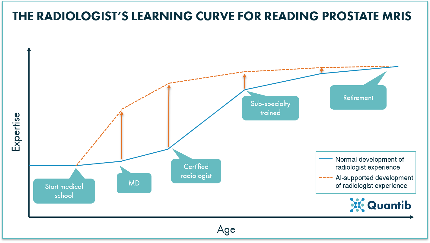
Figure 1: Without AI a radiologist starts his learning curve when he/she starts medical school (blue solid line). The gained expertise for reading prostate MRIs will increase in time, with the strongest slope during sub-specialty training. Implementing AI software to support radiologists will allow them to use highly specialized knowledge earlier on in their careers.
What can AI do to help the radiologist?
There are many ways in which AI can support radiologists with the challenging task of reading prostate MRIs. Providing an all-round software package offering a range of assisting AI functionalities will make the lives of radiologists easier and, most likely, more efficient, as performing daily tasks with well-designed and comprehensive solutions can provide an efficiency boost to day-to-day task.
Firstly, AI software can avoid the radiologists’ need to do their assessment divided over many places. For example, a software, thank to their AI algorithms, can automatically, and in seconds, determine prostate volume and PSA density, which not only eliminates the imprecise ellipsoid approach from the process, but also saves the radiologist time.
Moreover, the software can also provide automated analysis that assesses multiple sequences in one go. Such information can be presented in an intuitive heatmap making it easier to visualize the combined information and highlighting possible regions of interest.
Furthermore, AI software can streamline the workflow, for example, by providing step-wise guidance for the determination of the PI-RADS score. Each essential input will be requested by the software, guiding the user through the PI-RADS guidelines and providing valuable support to get to a final PI-RADS scoring.
With all support AI software can provide, such a program can be specifically useful for starting radiologists to improve their reading skill early on. Nonetheless, prostate MRI can be challenging field for radiologists of all levels of expertise, especially if we take into account the increase in workload previously mentioned. Stress and mental fatigue can lead to inter- and intra- observer variability which can negatively impact the efficiency of the radiologist workflow. However, functioning as a second opinion, the automated calculations and analysis AI provides can reduce that variability.
The automated generation of a standardized report can be a significant asset for multidisciplinary team meetings (MDTs). The report can include key images, which otherwise may take additional time to collect and save. In case of some PACS vendors, this time may even have to be spent during MDT, as images need to be looked up on the spot. Additionally, easily understandable graphs and metrics provide valuable context for decision making. Consequently, the amount of time radiologists need to prepare for these meeting will be reduced and the meetings themselves will be more time-efficient.

How can AI minimize the urologist’s challenges associated with prostate MRIs?
AI solutions for prostate MRI reading may result in a more efficient information exchange between radiologists and urologists.
As an AI software can reduce the amount of time it takes radiologists to assess the images and report their findings, the turn around time for the radiology exam may be shortened as well as results which translates into less waiting time for the patient to receive the outcome of their diagnosis trajectory.
Moreover, the decrease in inter- and intra- observer variability will make the results easier to interpret by a urologist and easier for him to make better informed decisions about the next steps in the diagnostic process of their patients. During communication with the patient, the AI-generated report may also provide an easy-to-understand base to support the urologist explaining the situation and treatment option.
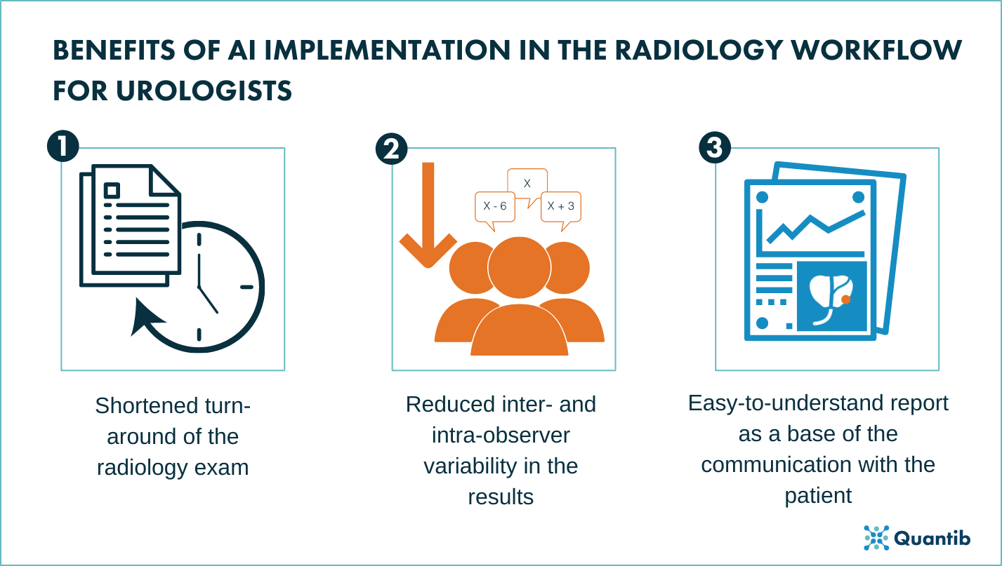
Figure 2: Key benefits of AI implementation in the radiology workflow for urologists.
Conclusion
These many challenges, from the heavier workload to the non-intuitive interfaces, present a significant risk to the mental health of the professionals involved in the clinical pathway for PCa diagnosis. As these challenges can lead to continuous stress and burnout, it is essential to tackle these challenges to avoid a decrease in quality of patient care.
Although recent research7 shows there is always room for improvement when it comes to the application of AI to prostate MRI, incorporating AI into the radiology workflow may solve many challenges starting today. Therefore, AI driven solutions for radiology can play a key role in improving the day-to-day conditions these professionals encounter and advance the quality of patient care.
Are you curious to know how Quantib supports the prostate MRI workflow? Then learn more about Quantib®Prostate, our AI driven solution for advancing the clinical pathway for prostate cancer (PCa) diagnosis.
Bibliography
- Ahmed, H. U. et al. Diagnostic accuracy of multi-parametric MRI and TRUS biopsy in prostate cancer (PROMIS): a paired validating confirmatory study. The Lancet 389, (2017).
- Andrew B. Rosenkrantz. MRI-Targeted or Standard Biopsy for Prostate-Cancer Diagnosis. New England Journal of Medicine (2018) doi:10.1056/nejmoa1801993.
- Mottet, N. et al. EAU Guidelines. (2020).
- Aprikian, S. et al. Improving ultrasound-based prostate volume estimation. BMC Urology 19, (2019).
- Rodriguez, E., Skarecky, D., Narula, N. & Ahlering, T. E. Prostate Volume Estimation Using the Ellipsoid Formula Consistently Underestimates Actual Gland Size. Journal of Urology 179, (2008).
- Rosenkrantz, A. B. et al. The learning curve in prostate MRI interpretation: Self-directed learning versus continual reader feedback. in American Journal of Roentgenology vol. 208 W92–W100 (American Roentgen Ray Society, 2017).
- Penzkofer, T. et al. ESUR/ESUI position paper: developing artificial intelligence for precision diagnosis of prostate cancer using magnetic resonance imaging. European Radiology (2021) doi:10.1007/s00330-021-08021-6.