Artificial intelligence (AI) is redrawing the healthcare landscape and neurology is no exception to this growing trend. Not without reason, because it can offer many benefits, both in the realm of neurological research, as well as in diagnosis and therapeutic interventions. But what is AI and how can neurologists use it to their advantage? How can AI-based algorithms help neurologists and their patients? In this article, we will explore five areas of neurology where AI is currently playing a role in improving healthcare.
Let's start with some AI basics
AI covers the field of computer science that is focused on simulating intelligent human behavior and computational processes within the brain. Other terms, such as “machine learning” and “deep learning” are sometimes used as synonyms of AI. Yet, these are subfields of the complete field of artificial intelligence.
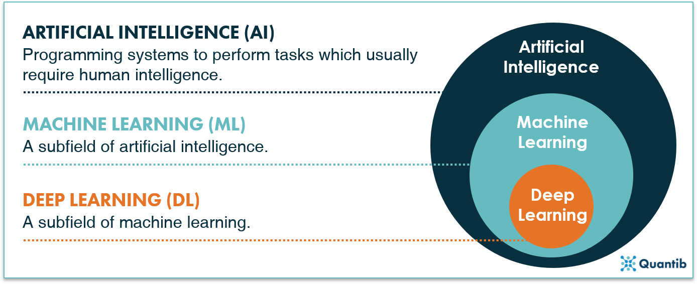 Figure 1: Artificial intelligence covers all programming systems that can perform tasks which usually require human intelligence. Machine learning and deep learning have the same capabilities, but use specific methods with machine learning being a subfield of AI and deep learning being a subfield of machine learning.
AI is probably the most talked about technology, but AI means different things to different people. Usually, people think of algorithms that can learn patterns from large datasets in order to be able to recognize these patterns later in new data. These can be both machine learning and deep learning algorithms.
Read our Ultimate Guide to AI in radiology and discover how all these algorithms work.
AI in neurology
AI applications for neuro-oncology
The field of neuro-oncology knows many challenges for which AI can offer support. This can be seen in the example of brain tumor assessment and diagnosis. Researchers from Heidelberg University Hospital and the German Cancer Research Center have trained a machine learning algorithm using approximately 500 magnetic resonance imaging (MRI) scans of patients suffering from brain tumors. Using volumetric tumor segmentation as a ground truth, the resulting algorithm was able to detect and localize brain tumors automatically on the MRI scans1. Such techniques can be of great value in, for instance, accurate diagnoses, and can also assist in tracking tumor therapy response in a repeatable and objective way.
Another application of AI in neuro-oncology is outcome prediction. Emblem et al. developed a machine learning algorithm (a support vector machine) using MRI-based blood volume distribution data to predict preoperative glioma survival. Results have shown that the algorithm was able to estimate survival between 6 months and 3 years, with even higher accuracy than experts in aggressive glioma cases2.
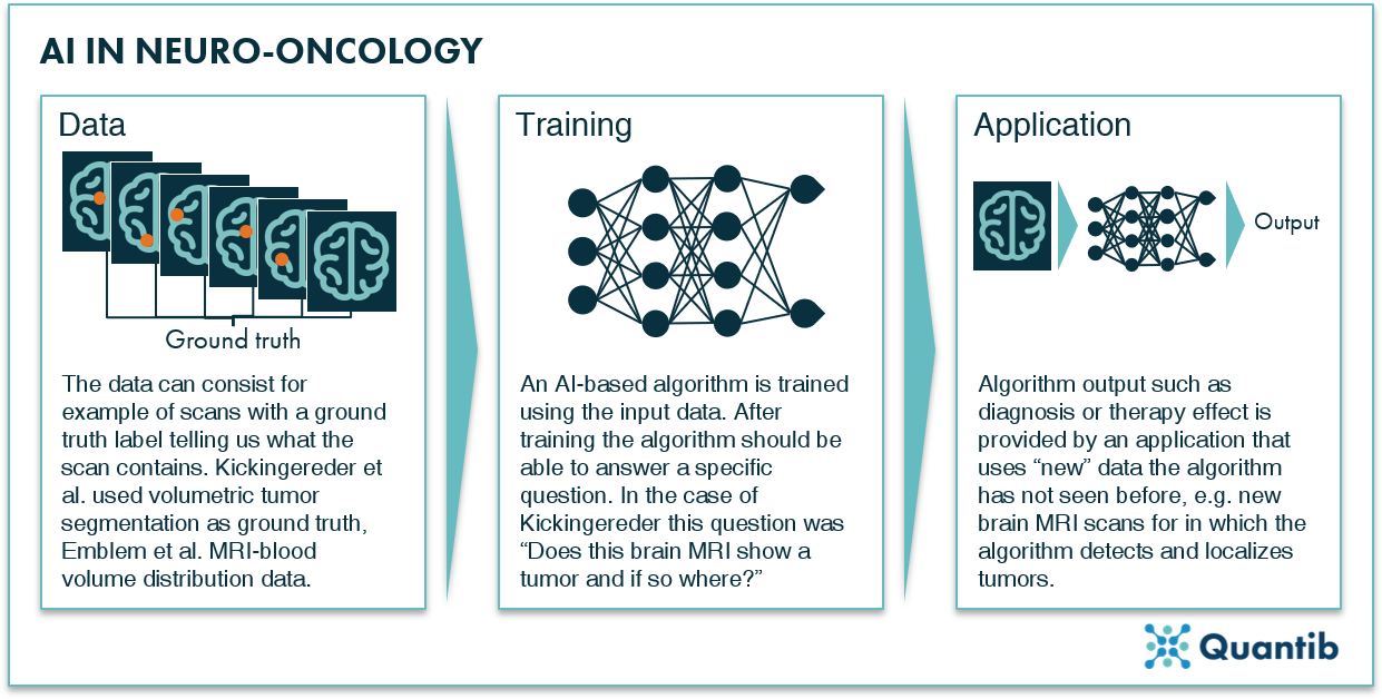 Figure 2: AI in neuro-oncology: data with ground truth are used for algorithm training. After training, the algorithm will be able to provide an output (answer a specific question, such as “does this brain MRI show a tumor and if so where?”) based on new data.
AI applications for neurodegenerative diseases
AI has been helpful in the field of neurodegeneration which includes Parkinson’s, Alzheimer’s, and Amyotrophic Lateral Sclerosis (ALS). A nice overview of the application of AI kinematic data for the diagnosis and assessment of Parkinson’s disease was published in 20193. Several (types of) algorithms were compared for different applications (diagnosis vs assessment) along different body sites. The authors discussed a wide range of methods and corresponding results, which only covered a limited part of all possible applications of AI in the area of Parkinson’s disease, as it did not include the evaluation of MRI data, speech recordings, or AI-support in a search for improved medication, just to name a few3.
Similar research around AI and Alzheimer’s disease has been performed in the past. AI has been applied to speech recordings4, cognitive tests scores5, and, perhaps most extensively, to neuroimaging, as it offers a wide range of AI-based image analysis options. For example, through analyses of brain MRIs. AI algorithms can automatically measure biomarkers of Alzheimer’s disease, including the rate of brain atrophy6. If large datasets are available, AI is able to compare a patient’s MRI results to a normative value, and thus provide information on whether a patient’s biomarker is within a normal range or deserves extra attention7.
Discover Quantib's FDA cleared and CE-marked AI-driven software for brain atrophy and WMH quantification.

Different approaches in AI can also be deployed to analyze data other than medical images. Researchers used sophisticated AI-based text search to sift through published literature on RNA-binding proteins that were previously linked to ALS8. They determined semantic features typical for ALS-related proteins. Subsequently, this information was used to scan other literature for proteins that are described using the same semantic features. Through this effort, the researchers singled out five RNA-binding proteins previously unlinked to ALS (for those interested in the details: specifically, the proteins hnRNPU, Syncrip, RBMS3, Caprin-1, and NUPL2 all showed significant alterations in ALS patients compared to controls)8.
AI applications in the neurovascular field
Many applications of AI in the field of neurovascular disorders have been researched and even brought to market as software packages over the past few years. For instance, AI can aid the diagnosis of stroke in various ways. AI-based assessments of CT scans of hemorrhagic patients allow, for example, the automated lesion segmentation of hemorrhagic infarcts9 or the automated detection and quantification of hemorrhagic expansion10. Additionally, AI has shown the ability to detect early warning signs of ischemia on CT images or to measure the extent of early ischemic changes by automated determination of the ASPECT score11. One last example of AI’s value in stroke diagnosis is the measurement of the time of ischemic stroke onset. These are just a few examples, but many more are available, so in case of curiosity, a quick Pubmed search might give you more ideas.
Outside the AI application in the neurovascular imaging analysis domain, equal success can be expected in other parts of the neurovascular field. For example, Labovitz and colleagues, from the Montefiore Medical Center and AiCure located in New York City, demonstrated that AI can aid in patient monitoring, particularly in regard to medication. In Labovitz et al.’s study, the AI platform was installed on the patient’s mobile device where the AI algorithms were able to visually identify the patient, the medication, and the confirmed ingestion of that medication. This study also found that patients receiving this real-time monitoring had increased adherence to their prescription regimen12. Thus, deploying a patients’ smartphone by installing AI-based apps that can monitor one’s behavior and treatment adherence can help improve therapeutic outcomes. This may seem like quite a simple and unadvanced example, but one can imagine that the difference it may be able to make in people’s lives could be substantial.
AI applications for traumatic brain injury
Applying AI to traumatic brain injury (TBI) has also been fruitfully researched over the past years. The main goal of intensive care in case of TBI is to mitigate the impact of secondary brain injury following the first insult by controlling factors such as intracranial and cerebral perfusion pressure. In 2019, researchers from Finland published a study about the use of AI in cases of TBI. These researchers developed an algorithm that could predict 30-day mortality for TBI patients. This algorithm was trained only on a few main variables (intracranial pressure, cerebral perfusion pressure, and mean arterial pressure). A secondary algorithm that utilized the motor and eye movement components of the Glasgow Coma Scale was also trained. Remarkably, with these few variables, the AI was able to accurately discriminate between survivors and non-survivors over 80% of the time13.
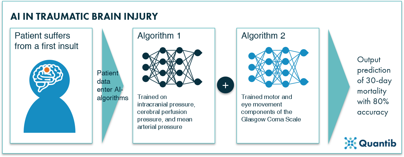 Figure 3: AI in traumatic brain injury: Raj et al. trained two algorithms which, combined, can predict 30-day mortality with 80 percent accuracy in patients that suffered from TBI.
AI applications for spinal cord injury
Finally, applications of AI in neurology can be seen in cases of spinal cord injury. A study published in the journal Nature by researchers from the Battelle Memorial Institute and Ohio State University has recently shown that intracortically recorded signals can be linked in real time to muscle activation to restore movement in a paralyzed human. Researchers used a machine learning algorithm to decode the neuronal activity and control activation of forearm muscles through a custom-built electrical stimulation system14. This enabled a 24-year-old man who had previously lost function in his hands and arms due to spinal cord injury to once again grasp, manipulate, and release objects. This represents not only an important contribution to neuro-prosthetic technology but also has important and direct implications for people living with paralysis worldwide.
Using AI in neurology in the clinic
We have outlined several ways in which AI has shown promise in the field of neurology. Many algorithms still need to go through the important translation process from a research tool to clinically approved software, while some applications have already obtained a nod from the regulatory bodies for use in the clinic. All in all, AI in neurology proves to be a promising field of research, and, if you are interested in bringing artificial intelligence software to your neurology practice, there are quite a few options available on the market. Just make sure to select software that has been approved for clinical use.
Bibliography
- Kickingereder, P. et al. Articles Automated quantitative tumour response assessment of MRI in neuro-oncology with artificial neural networks : a multicentre , retrospective study. 1–13 doi:10.1016/S1470-2045(19)30098-1
- Emblem, K. E. P. et al. Machine Model for Preoperative. Radiology 275, (2015).
- Belić, M. et al. Artificial intelligence for assisting diagnostics and assessment of Parkinson’ s disease — A review. 184, (2019).
- Chien, Y., Hong, S., Cheah, W., Yao, L. & Chang, Y. An Automatic Assessment System for Alzheimer’ s Disease Based on Speech Using Feature Sequence Generator and Recurrent Neural Network. 1–10 (2019). doi:10.1038/s41598-019-56020-x
- Hughes, O. Using AI assessment to tackle dementia in ultra-early stages. (2019). Available at: https://www.digitalhealth.net/2019/09/using-ai-assessment-tackle-dementia-ultra-early-stages/.
- Blamire, A. M. Progress in Nuclear Magnetic Resonance Spectroscopy MR approaches in neurodegenerative disorders. Prog. Nucl. Magn. Reson. Spectrosc. 108, 1–16 (2018).
- Vinke, E. J. et al. Neurobiology of Aging Normative brain volumetry derived from different reference populations: impact on single-subject diagnostic assessment in dementia. 84, (2019).
- Bakkar, N. et al. Artificial intelligence in neurodegenerative disease research: use of IBM Watson to identify additional RNA ‑ binding proteins altered in amyotrophic lateral sclerosis. Acta Neuropathol. 135, 227–247 (2018).
- Scherer, M. et al. Development and Validation of an Automatic Segmentation Algorithm for Quantification of Intracerebral Hemorrhage. 1–8 (2016). doi:10.1161/STROKEAHA.116.013779
- Masaki Nagamine, Paul Kohanteb, Wengui Yu, Michelle Bardis, Te-Chang Wu, Lydia Su, Daniel Chow, Peter Chang, Jeon Chen, J. S. Abstract WP395: Detection of Hemorrhagic Expansion With AI. Stroke (2020).
- Takahashi, N. et al. Computer-aided detection scheme for identification of hypoattenuation of acute stroke in unenhanced CT. Radiol. Phys. Technol. 5, 98–104 (2012).
- Labovitz, D. L., Shafner, L., Gil, M. R., Virmani, D. & Hanina, A. in Patients on Anticoagulation Therapy. 48, 1416–1419 (2018).
- Raj, R., Luostarinen, T., Pursiainen, E., Posti, J. P. & Takala, R. S. K. Machine learning-based dynamic mortality prediction after traumatic brain injury. 1–13 (2019). doi:10.1038/s41598-019-53889-6
- Bouton, C. E. et al. in a human with quadriplegia. Nature 1–13 (2016). doi:10.1038/nature17435