Artificial intelligence (AI) is conquering the world and the field of healthcare is no exception to this revolution. Even though its clinical implementation is still cautious, many are convinced AI is influencing our lives and will do so increasingly in the near future. AI-based medical imaging assessments, AI-guided surgery, AI-steered hospital bed utilization, are just a few examples of AI implementation to healthcare as an industry.
With this article, we want to provide insights into some of the most recent developments of AI. However, as healthcare includes a vast amount of disciplines, we will be focusing on the field of urology for a couple of reasons.
In the first place, urology is a very diverse specialization with many different types of interactions with patients, colleagues and machines, it makes for an interesting area for AI implementation.
Secondly, the field of urology overlooks multiple cancers that are part of the top 10 most suffered cancer types by men in 2020 worldwide1, such as prostate, bladder and kidney cancer. For that reason, any advancements in urology can have a big impact in a lot of patients lives.
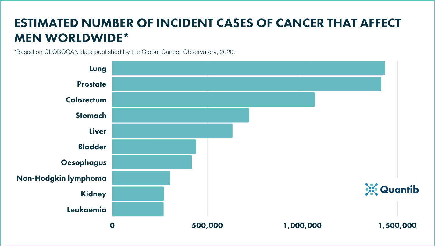
Figure 1: Table chart of the highest incident cases of cancer suffered by men, all ages in 20201.
The field of AI in urology
To provide you with the right context for understanding the developments in AI in urology, we developed a framework showing the amount of research going on in the different fields.
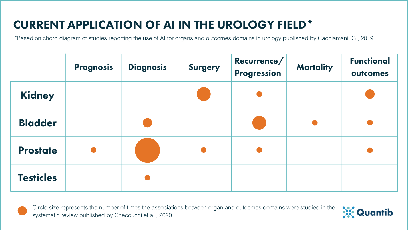
Figure 2: Summarized overview of the studies reporting the use of AI for organs and outcome domains in urology based on “Artificial intelligence and neural networks in urology: current clinical applications”2.
As the figure shows, the application of AI in urology has been widely investigated. A few areas are more popular than others, for example, diagnosis in prostate with solutions to support prostate MRI reading; surgery in kidney, and recurrence/progression in bladder, with AI to support the prediction of recurrence-free probability in patients with superficial urothelial bladder cancer. However, the figure also shows the potential of AI applications would the research expand and deepen.
AI developments in prostate cancer
Due to the incidence of prostate cancer and the predominant research done in the subspecialty of prostate oncology, we will focus on discussing some of the many applications that are increasingly part of the prostate cancer pathway, covering examples in the areas of histopathology and prostate MRI.
AI for tumor detection in histopathology
Applied to prostate cancer detection, a Computer Aided Diagnosis (CAD) system normally receives the histopathological images in its raw state and processes them for a diagnostic result. AI and its developments are the base for the growth of these CAD systems3.
While there is still improvement to be done in CAD systems, research has shown that “histopathology images may enhance the early diagnosis and treatment of prostate cancer patients through providing functional and morphological data about the prostate”3. The same research also showed a prevalent use of deep learning (DL) and machine learning (ML) techniques in histopathology.
The use of CAD in this discipline is focused on reducing human errors to improve the accuracy of the diagnosis by, for example, improving the precision and quality of histopathological images, segmenting different parts of the tumor and even classifying diseases. For example, support vector machine (SVM) algorithms for classification of histopathology images can reach accuracy of 95% for automatically differentiating between artefacts and glands and close to 90% in detecting pathological deviations versus benign tissue3.
Research applied to histopathological diagnosis4 computed an AI tool using Artificial Neural Networks (ANNs) that achieved an “area under the ROC curve of 0.997 for distinguishing between benign and malignant biopsy cores” which is said to be comparable to the accuracy that can be expected from an expert in prostate pathology.
AI for tumor grading in histopathology
When it comes to grading of prostate cancer, AI has been proven to reduce inter-variability between pathologists’ gradings, therefore increasing the certainty of prostate cancer diagnosis. Previous research5 proved that the consensus between pathologists (not subspecialized) that used AI is higher than the one between 3 uropathologists that did not use any AI support (0.872 vs. 0.799 respectively).
The same author later led, alongside a lot of other professionals, a worldwide histopathology competition, known as PANDA challenge6, in order to advance the reproducibility of AI algorithms for Gleason grading, as this has been seen as one of the major setbacks when it comes to the clinical implementation of such AI.
The results of the challenge further supported the previous results as AI algorithms presented higher agreement with expert subspecialty pathologists (0.862 and 0.868 for US and European datasets respectively) than the agreement reached between general pathologists and subspeciality pathologists. This is quite an important finding for the field as it confirms that AI could help alleviate the variability that is commonly associated with Gleason grading6.
Going one step further, this challenge found that algorithms generally missed less cancers than pathologists did (1% and 1.9% vs 1.8% and 7.3% in European and US datasets respectively)6. This confirms the value and the potential of AI when it comes to reducing undegrading and underdiagnosing prostate cancer.
AI for standardization in histopathology
Although AI systems for histopathology need to develop further in order to ensure a successful application to clinical practice, research has been promising.
One of the current difficulties is the lack of standardization in processing protocols for the histopathological images. Researchers7 mention how AI is welcomed and encouraged as a tool in prostate pathology because it will help standardize diagnostic grading and improve quality control which will, in turn, provide the urologist with more accurate assessments.
Achieving protocol standardization will also help overcome another difficulty CAD systems face in histopathology: the small availability of quality training datasets, especially of less common variants or benign cases of prostate cancer7.
AI for prostate MRI
A recent position paper8 reviews different AI developments related to prostate MRI and explores their bias. For example, AI tools to support diagnosis in radiology are said to deliver a high percentage of false positives in order to achieve high sensitivity for the detection of prostate cancer. This percentage can be as high as 50% in bpMRI8,9.
AI, as a radiological diagnosis support tool, has been suggested to overcome this high rate of false positives8. Such support tools need the radiologist’s approval of the automated AI detected lesions. Semi-automatic systems are a widely adopted approach by solutions that are currently available.
Discover Quantib®Prostate, our radiology AI software for optimizing the prostate MRI reading process.
![]()
A broad incorporation of AI in the field is thought to be able to decrease the financial burden of prostate cancer diagnosis and further treatment4.
While some researchers8 provide a critical note towards the current implementation of AI in prostate MRI, the research community is experimenting with approaches that may be increasingly valuable for clinical practice. For example, researchers developed a neural network that is able to simultaneously detect and grade cancer tissue10. A U-net was trained using MRI slices as input and Gleason gradings as output covering multiple aspects of the diagnostic process. An interesting approach, that could, with sufficient performance numbers, add substantial value in clinical practice.
AI for risk stratification
When it comes to the current classification system for Prostate Cancer Risk Stratification there is one main challenge to face, which is the fact that none of the risk categories look into recurrence as a factor. Deep learning provides a promising tool to look into recurrence as well as the other risk categories. Researchers looked into recurrence prediction by assessing the accuracy of two different convolutional neural networks (one focused on the detection of nuclei and the other on the classification of patches around the nuclear centers) which resulted in having an end yield of 0.81 AUC. This showed the potential of deep learning algorithms for predicting recurrence4,11.
Taking all these into account, it is safe to assume the impact and the importance of the possible application of AI to the field of prostate cancer.
Conclusion
The academic community has delivered impressive research and proven the multiple benefits of AI application to the urology field, and more specifically, the prostate cancer diagnostic workflow. We look forward to seeing more and more of these developments being introduced into the clinical practice.
Discover other AI developments for bladder cancer, kidney diseases or urologic surgery support.
![]()
Bibliography
- Global Cancer Observatory. Cancer today: Estimated number incident cases and deaths worldwide, males, all ages. International Agency for Research on Cancer (2021).
- Checcucci, E. et al. Artificial intelligence and neural networks in Urology: Current clinical applications. Minerva Urologica e Nefrologica vol. 72 49–57 (2020).
- Ayyad, S. M. et al. Role of ai and histopathological images in detecting prostate cancer: A survey. Sensors vol. 21 (2021).
- van Booven, D. J. et al. A systematic review of artificial intelligence in prostate cancer. Research and Reports in Urology vol. 13 31–39 (2021).
- Bulten, W. et al. Artificial intelligence assistance significantly improves Gleason grading of prostate biopsies by pathologists. Modern Pathology 34, (2021).
- Bulten, W. et al. Artificial intelligence for diagnosis and Gleason grading of prostate cancer: the PANDA challenge. Nature Medicine (2022) doi:10.1038/s41591-021-01620-2.
- Egevad, L. et al. The emerging role of artificial intelligence in the reporting of prostate pathology. Pathology vol. 53 565–567 (2021).
- Penzkofer, T. et al. ESUR/ESUI position paper: developing artificial intelligence for precision diagnosis of prostate cancer using magnetic resonance imaging. European Radiology (2021) doi:10.1007/s00330-021-08021-6.
- Schelb, P. et al. Classification of Cancer at Prostate MRI: Deep Learning versus Clinical PI-RADS Assessment. Radiology 293, 607–617 (2019).
- Vente, C. de, Vos, P., Hosseinzadeh, M., Pluim, J. & Veta, M. Deep Learning Regression for Prostate Cancer Detection and Grading in Bi-Parametric MRI. IEEE Transactions on Biomedical Engineering 68, (2021).
- Kumar, N. et al. Convolutional neural networks for prostate cancer recurrence prediction. in (eds. Gurcan, M. N. & Tomaszewski, J. E.) (2017). doi:10.1117/12.2255774.